Difference between revisions of "ExamplesDynamic/it"
(Created page with "*'''Due modelli nell'Articolatore A''' per Artex *'''Due modelli nell'Articolatore S''' per Sam o Adesso *'''Due modelli nell'Articolatore P''' per Protarevo- Kavo *'''Due mod...") |
(Created page with "Per maggiori informazioni su come creare progetti in Exocad, visitare la pagina [http://wiki.exocad.com/wiki/index.php?title=DentalDB_Module Exocad Wiki!]!") |
||
| Line 23: | Line 23: | ||
*'''Due modelli nell'Articolatore D''' per Denar by Whipmix | *'''Due modelli nell'Articolatore D''' per Denar by Whipmix | ||
| − | + | Per maggiori informazioni su come creare progetti in Exocad, visitare la pagina [http://wiki.exocad.com/wiki/index.php?title=DentalDB_Module Exocad Wiki!]! | |
Here is the project definition for our demo project; we have used the Articulator Protarevo by Kavo: | Here is the project definition for our demo project; we have used the Articulator Protarevo by Kavo: | ||
Revision as of 07:27, 23 June 2017
In questa pagina è mostrata e spiegata la procedura guidata da seguire per scansionare 2 modelli in occlusione con 3 monconi (2 superiori e 1 inferiore) utilizzando il Modulo Articolazione Dinamica di Open Technologies.
Il Modulo Articolazione Dinamica permette all'utente di trasferire l'occlusione realizzata in laboratorio con un articolatore fisico nel suo equivalente virtuale in Exocad. Gli articolatori supportati in Exocad, per i quali possiamo fornire posizionamento esatto, sono Artex, Protarevo Kavo, Sam, Bioart A7 e Denar by Whipmix.
Quando si abilita il Modulo Articolazione Dinamica, si fornisce all'utente un set di 6 Piattelli Splitcast ed un Oggetto di Calibrazione. Vedere Accessori- Modulo Articolazione Dinamica.
Prima di usare questo Modulo per la prima volta è necessario calibrare gli assi dello scanner per lavorare in linea con la simulazione dell'occlusione. Per imparare a calibrare, visitare la pagina Calibrare lo scanner.
QUESTO E UN MODULO DI LICENZA AGGIUNTIVO DI OPEN TECHNOLOGIES: RICHIEDI AL TUO DISTRIBUTORE COME OTTENERLO!!
Contents
Definizione del progetto in Exocad
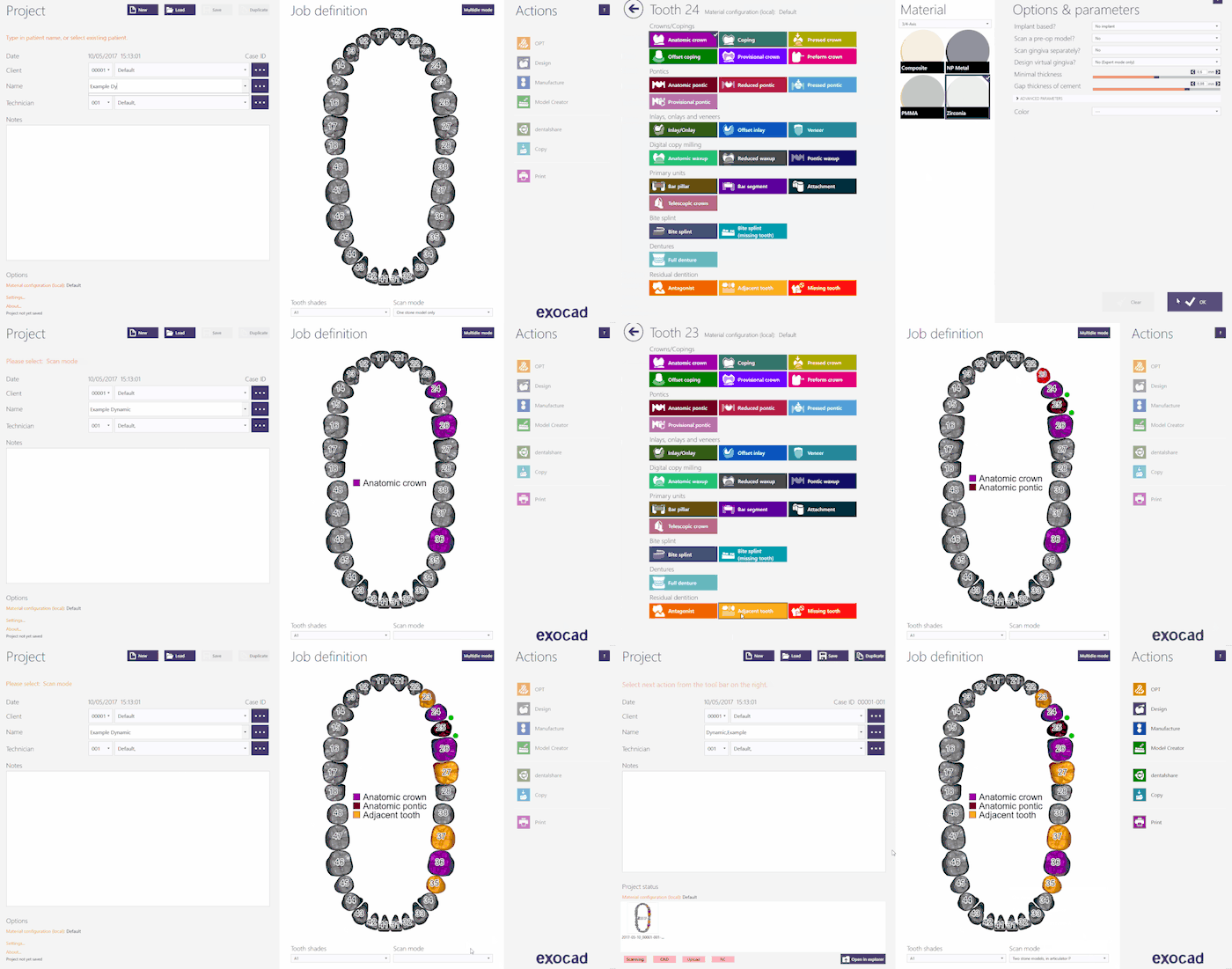
Per creare questo progetto in Exocad, cliccare sull'icona DentalDB sul desktop, aprendo così il Gestore Progetti.
Inserire le informazioni del progetto, il tipo di ricostruzione che dovrà essere modellata e i parametri aggiuntivi. Per questo tipo di progetto assicurarsi che la Modalità Scansione sia impostata su uno degli Articolatori virtuali di Exocad disponibili; scegliere tra:
- Due modelli nell'Articolatore A per Artex
- Due modelli nell'Articolatore S per Sam o Adesso
- Due modelli nell'Articolatore P per Protarevo- Kavo
- Due modelli nell'Articolatore B per Bioart's A7
- Due modelli nell'Articolatore D per Denar by Whipmix
Per maggiori informazioni su come creare progetti in Exocad, visitare la pagina Exocad Wiki!!
Here is the project definition for our demo project; we have used the Articulator Protarevo by Kavo:
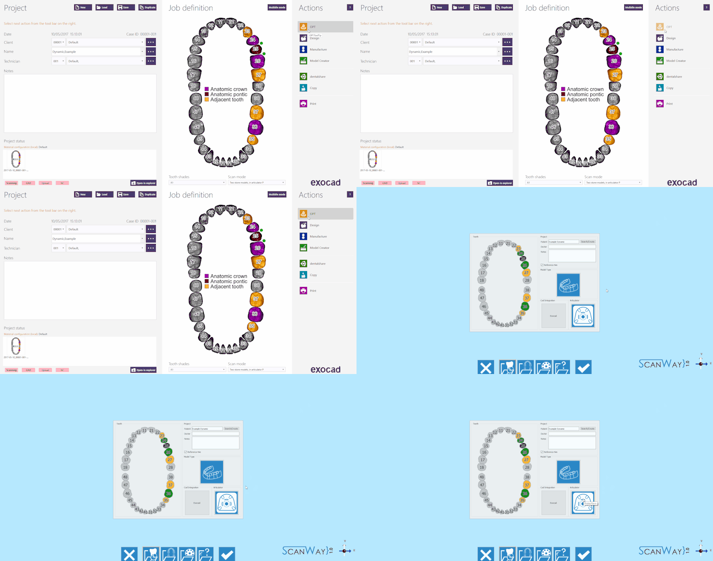
Once the project has been defined just click OPT in the Actions section to launch the scanning software.
The first thing the scanning software shows is the project created in Exocad. Check if the articulator selection is correct, if so click the Accept Button ![]() .
.
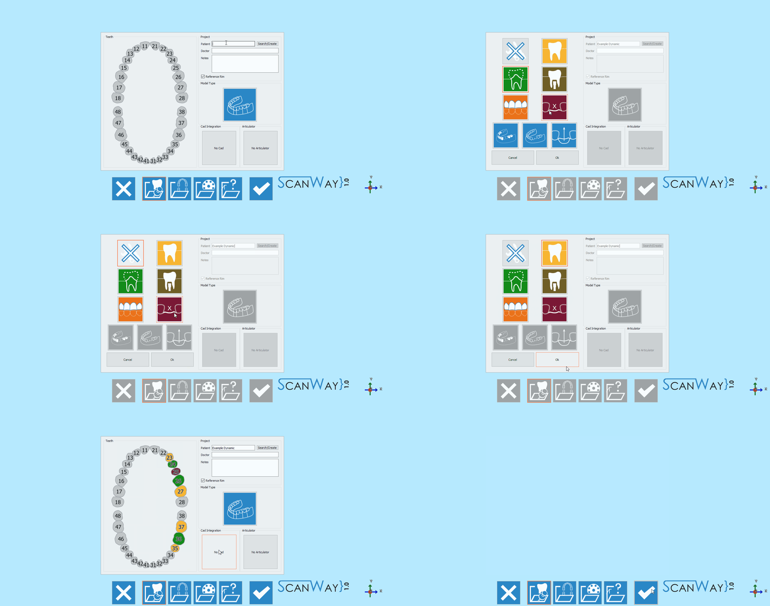
Project definition starting from ScanWay
All projects can be defined in the scanning software as well, regardless the CAD that will be later on used to design.
Launch the scanning software double-clicking on the ScanWay icon on your desktop. The Welcome Page will open; to create the project click on the first icon.
For detailed information on how to define a project visit the specific page: Create a New Project.
For this project, take particular care in the articulator selection. If the articulator gets wrongly defined, the virtual articulation won't be realistic and useful.
Also, remember to set Exocad as CAD platform, since this module is specifically designed to work for Exocad's Virtual Articulator.
Here is the definition of our demo project:
Both the project definitions described lead to the same wizard, which will be now followed step by step.
Step 1: Articulator Scanning
The first step of any project that has been set to have both jaws, is the Articulator Scan.
In this specific case the Articulator has to be scanned. The scanned articulator needs to match the one selected in the project definition.
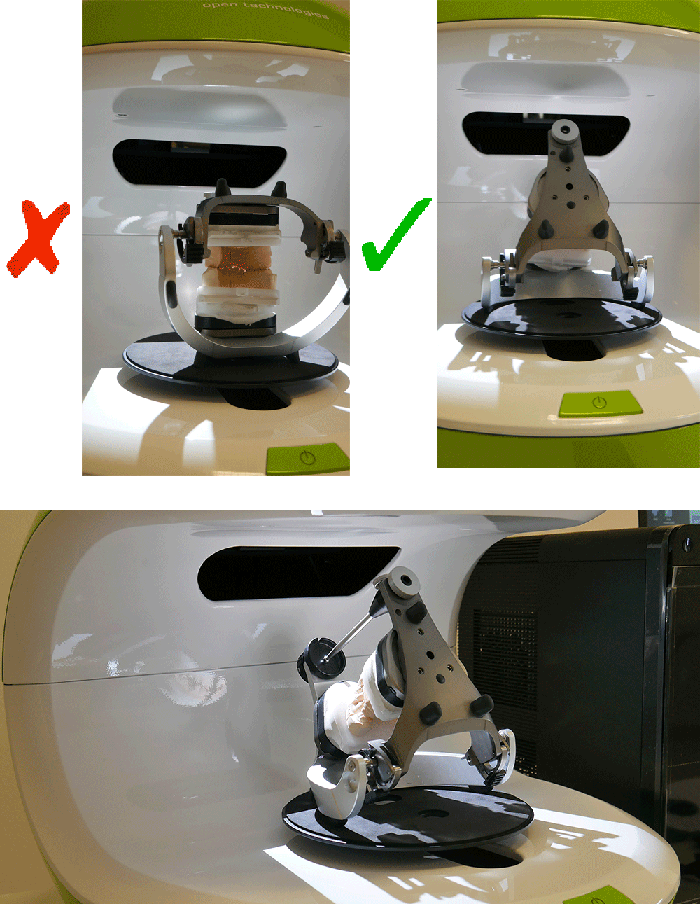
We suggest to scan the articulator tilting it backwards, to get as much information as possible. This is just a suggestion as not all articulators can be tilted.
In the live view of the scanning interface you can actually see that if the articulator is standing straight it will be difficult to acquire the occlusion properly.
Once the articulator has been placed in the scanner, the scan can be started with the Scan Button![]() . Once the scan is finished the result will be shown.
. Once the scan is finished the result will be shown.
For further information on the other available functions check the Scan Interface page.
Click ![]() to access the second step of the wizard.
to access the second step of the wizard.
Step 2: Lower Model
The second step of any project that has been set to have both jaws, is the Lower Model Scan. All steps, except for the articulator one, include two sub-steps: the actual scan and the edit of the acquired image.
Scan Step
Place the lower model on the splitcast base matching the selected articulator; for our demo case we used the Protarevo- Kavo Splitcast base.
Click the Scan Button![]() . Once the scan is finished the result will be shown.
. Once the scan is finished the result will be shown.
Click ![]() to access the second part of the Lower model step.
to access the second part of the Lower model step.
Edit Step
This step allows to edit the acquired image; for detailed information on all the available options in this step visit the Edit Tools page.
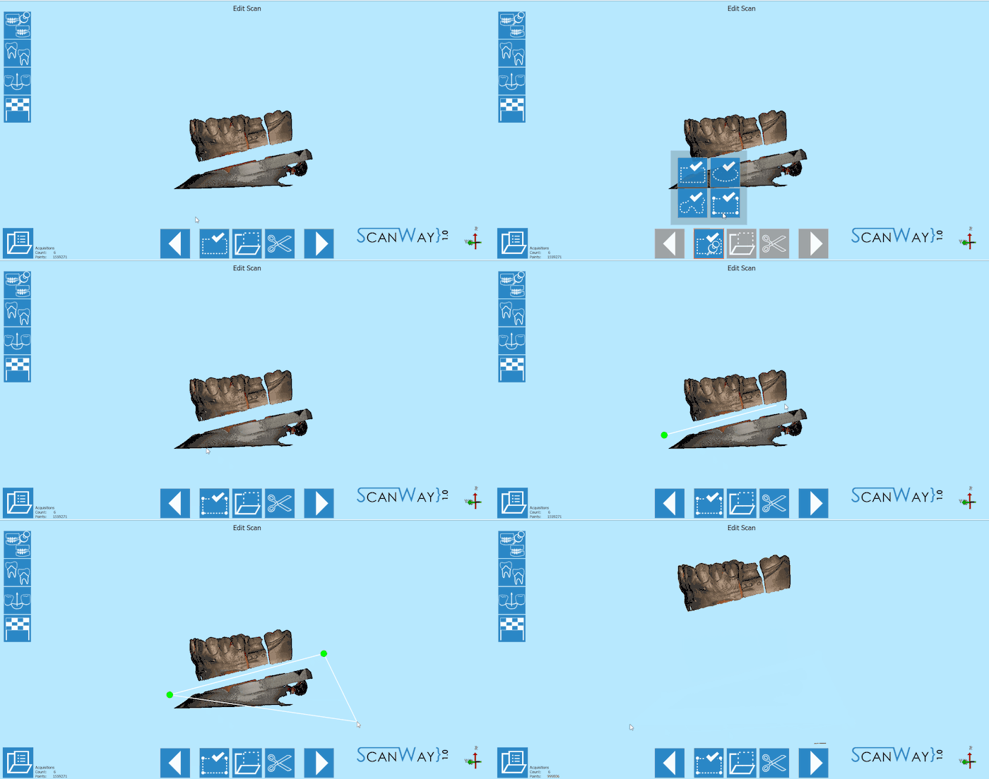
The image at this step can be both edited or trimmed. At this stage it is actually important not to edit too much the image or cutting big chunks of information, since it would make it more difficult for the software to calculate the automatic alignment of the object to its reference.
In this particular case, since the model is mounted in the articulator, it's not scanned on a perfectly horizontal plane, so to trim it faster we used the polygonal selection tool.
When you are satisfied with the result click ![]() to access the next wizard step.
to access the next wizard step.
Step 3: Upper Model
The upper model Scan, like the lower model, requires the user to undergo two steps.
Scan Step
Place the upper model on the splitcast base matching the selected articulator and click the Scan Button![]() . Once the scan is finished the result will be shown.
. Once the scan is finished the result will be shown.
Click ![]() to access the edit step for the Upper model.
to access the edit step for the Upper model.
Edit Step
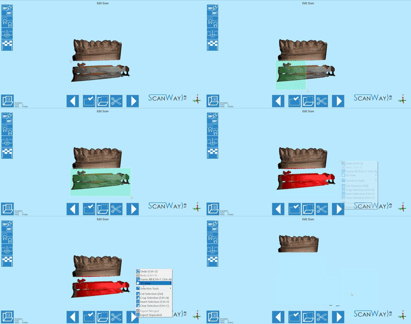
The upper model can still be edited as shown for the lower model. This time, we will edit the image using a different selection tool.
Step 4: Dies Scan
The steps necessary to successfully scan the dies vary depending on the scanning method that the user chooses to apply: the use of the multidie plate or the custom set-up.
With Multidie
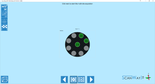
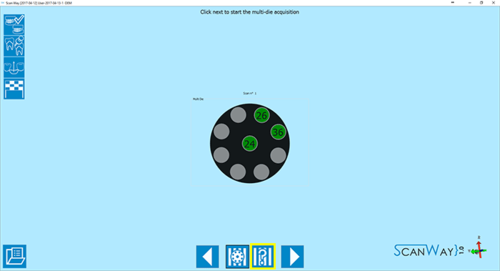
This method is applied by default by the software. It implies that the user will put the dies on the multidie plate in the position shown by the software, which will make each die immediately recognisable and cut the steps of the wizard.
- Definition
- As mentioned, the position of the dies in the multidie plate is pre-established by the software.
The multidie order is based on the Universal tooth numbering, starting from the last element of the first quadrant and following in a clock-wise order. Therefore the die that needs to be placed in the central element will always be the one closest to the last element of the first quadrant.
If there are more than 9 dies, a second definition step will be shown.
Click ![]() to access the multidie scan step.
to access the multidie scan step.
- Scan
- The multidie scan interface is similar to the other steps we just explained, except for the multidie reference that is kept as a reminder on the right side of the window, under the live view.
To proceed click the Scan Button . Once the scan is finished the result will be shown.
Click ![]() to access the edit step for the multidie scan.
to access the edit step for the multidie scan.
- Edit
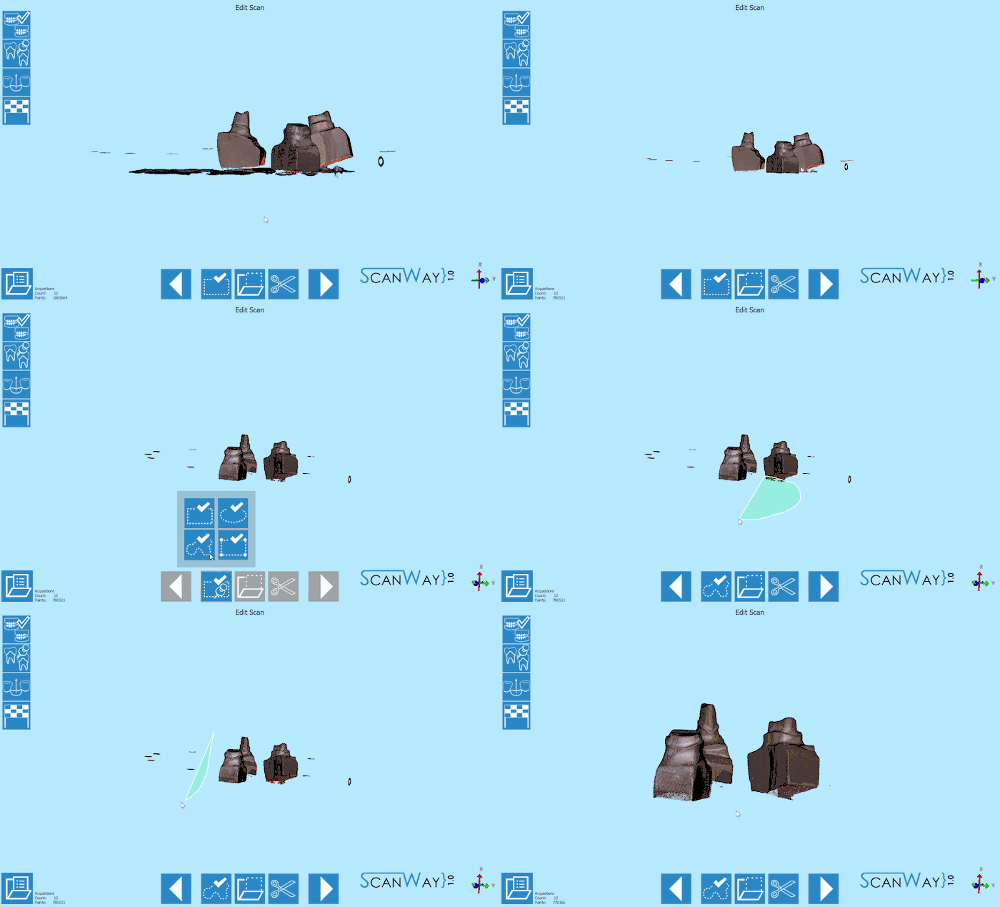
- The edit step offers the same tools we saw in previous steps. In this case, more than one tool may need to be applied.
When you are satisfied with the result click ![]() to access the next wizard step.
to access the next wizard step.
- Alignment
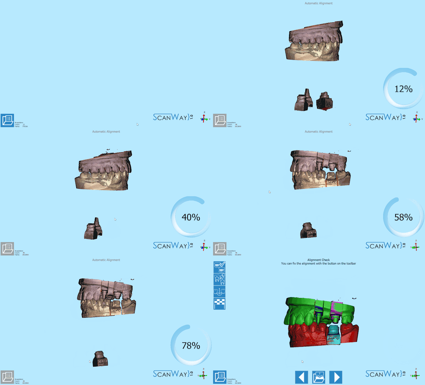
- After being edited the dies get aligned to their references. The result is then showed on the monitor. To learn how to change, fix or redo the alignment check the Alignment Interface page.
Custom Set-Up (Without Multidie)
The user can also decide to scan the dies in a custom order, for instance if he needs to scan the dies on the model base. To access the custom set- up definition, click on the ![]() icon when the software presents the multidie definition.
icon when the software presents the multidie definition.
- Definition
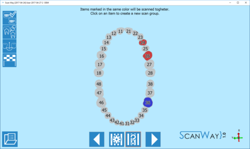
- The software proposes by default a unique scan group which means that, if not defined otherwise, the software will ask the user to scan all the stumps together. In this case I have created a second scan group, to divide the scan of the upper dies from the scan of the lower dies. Click
 to access the scan steps.
- Scan
- Depending on the number of scan groups created, the software will propose one or more scanning steps.
First, the software requires the user to insert the dies in the scanner and acquire the items of the first group.
As a reminder, it marks the items to be scanned together on the right of the window, under the live view.
Then it will ask the user to insert the item selected for the second group. Since the second scan is an individual die, there will be no reminder on the right. Click ![]() to continue.
to continue.
- Dies Identification
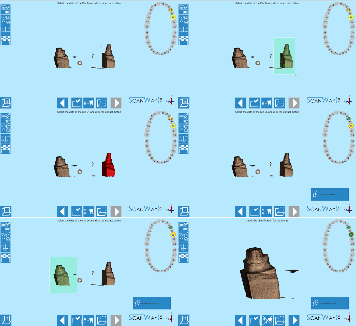
- When a scan group has more than one item, the user will be asked to separate each die from the others to correctly identify it.
This step occurs in between the scanning steps. To learn more on the identification of the dies visit the Scan Interface page. Click to continue.
- Edit
- If the scan group is formed by one individual die, there will be no identification step. Instead, the edit step will be presented. Click
 to continue.
- Alignment
- After being edited or identified the dies get aligned to their references. The result is then showed on the monitor. To learn how to change, fix or redo the alignment check the Alignment Interface page.
Step 5: Healthy and Pontics
At this stage the project has been scanned, unless the user wants to rescan the healthy and pontics. This step is infact a result of the scan of the scanned reference model, trimmed to exclude the parts that have been scanned individually (in this case, the dies). It sometimes happens that in the first model scan, the contact points on the healthy are not correctly recognised, which would require the user to rescan the model. Otherwise, just click ![]() to continue.
to continue.
The software will then propose a further edit step to allow the user to modify the healthy image. Proceed in the edit step as previously explained.
Step 6: Mesh Generation and Export
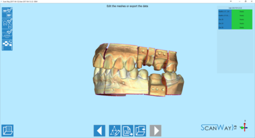
At this point the software immediately starts mesh generation. The meshes can be edited and exported individually or as a unique image. To learn more about mesh editing visit our Mesh Tools page.
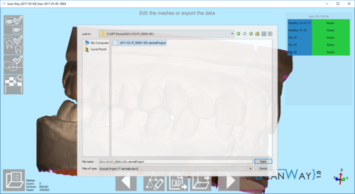
Click on the Export Button ![]() to export to CAD.
to export to CAD.
If the project has been started from Exocad, the CAD will automatically open and the design can be started immediately. Otherwise the software will ask the user how to export the file.
Since Exocad has been selected in ScanWay's project definition, the software will ask the user to find the folder containing the DentalProject file for this job.
