Difference between revisions of "ExamplesDynamic/ja"
(Created page with "{{Inline button|nextAction.png}} をクリックして、マルチダイスキャンステップに進みます。") |
(Created page with ";スキャン: マルチダイスキャンインターフェースはすでに紹介したほかのステップに似ていますが、相違点として、マルチダイの...") |
||
| Line 134: | Line 134: | ||
{{Inline button|nextAction.png}} をクリックして、マルチダイスキャンステップに進みます。 | {{Inline button|nextAction.png}} をクリックして、マルチダイスキャンステップに進みます。 | ||
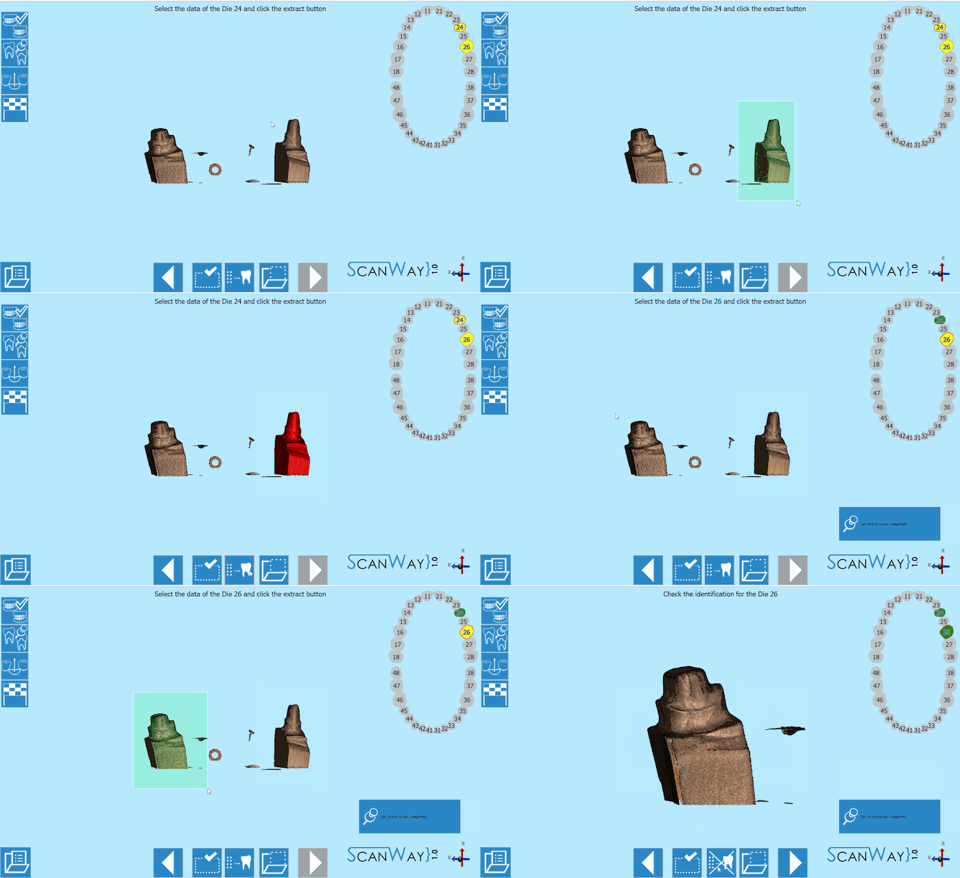
| − | ; | + | ;スキャン: マルチダイスキャンインターフェースはすでに紹介したほかのステップに似ていますが、相違点として、マルチダイのレファレンスがウインドウ右側のライブビューの下にリマインダーとして表示されます。<br /> 続行するには、'''スキャンボタン'''{{Inline button|scanAction.png}} をクリックします。スキャンが終了すると、結果が表示されます。 |
[[File:multidie-steps.png]] | [[File:multidie-steps.png]] | ||
Revision as of 08:50, 25 September 2017
このページでは、咬合する2つのモデルと3つのダイ(上顎2つと下顎1つ)を、オープンテクノロジーの ダイナミックアーティキュレーションモジュールを使ってスキャンする例を、ウィザードに従って説明します。
ダイナミックアーティキュレーションモジュールでは、技工所で実際の咬合器を使って設定した咀嚼位置の情報を、Exocadのバーチャル咬合器に移すことができます。Exocadで利用可能で、オープンテクノロジーが対応している咬合器には、Artex、Protarevo Kavo、Sam、Bioart A7、およびDenar by Whipmixがあります。
ダイナミックアーティキュレーションモジュールが有効化されると、ユーザーには4つのマウンティングプレートのセットと、1つのキャリブレーションキーが提供されます。付属品-ダイナミックアーティキュレーションモジュールを参照して下さい。
はじめてモジュールを使用する場合、スキャナーの軸のキャリブレーションを行い、咬合のシミュレーションに従って動作するように設定する必要があります。キャリブレーションの方法については、スキャナーのキャリブレーションのページを参照して下さい。
これは追加ライセンス用モジュールです。詳細はディーラーにお問い合わせ下さい
Contents
Exocadでプロジェクト定義を開始する
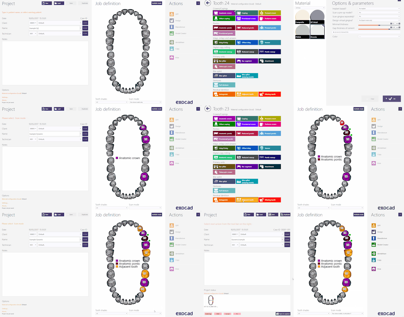
このプロジェクトをExocadで作成するには、デスクトップでDentalDBアイコンをクリックします。プロジェクトマネージャーが開きます。
プロジェクト情報、設計する修復の種類、その他のパラメーターを入力します。この種類のプロジェクトでは、Scan Mode(スキャンモード)が利用可能なExocadのバーチャル咬合器のいずれかに設定されていることを確認します。以下から選択します。
- Two stone models in Articulator A :Artexの場合
- Two stone models in Articulator S :SamまたはAdessoの場合
- Two stone models in Articulator P :Protarevo- Kavoの場合
- Two stone models in Articulator B :Bioart's A7の場合
- Two stone models in Articulator D :Denar by Whipmixの場合
Exocadでプロジェクトを作成する方法について詳しくは、Exocad Wiki!を参照して下さい。
今回のデモ用プロジェクトでは以下のプロジェクト定義を使用します。咬合器にはKavoのProtarevoを使用します。
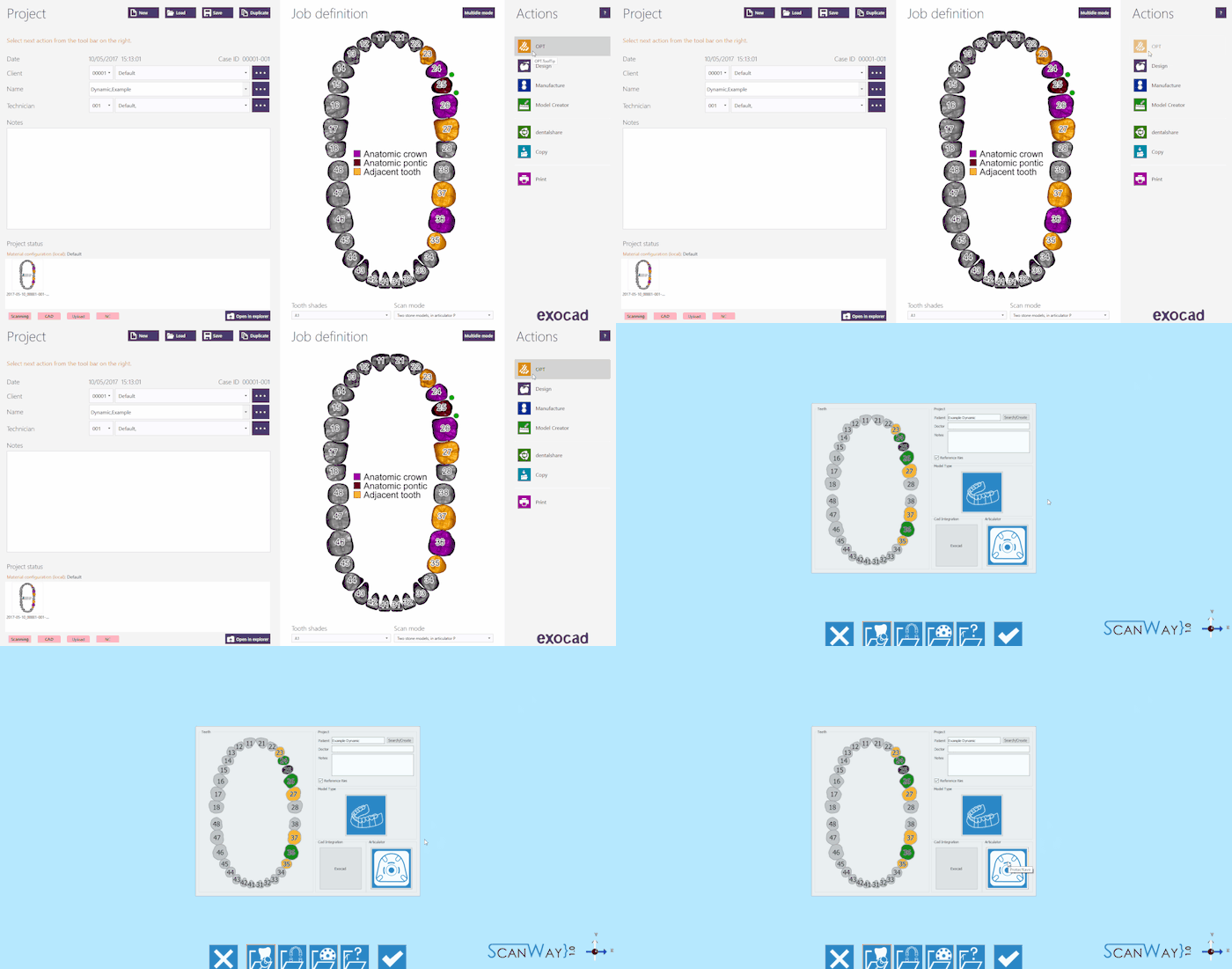
プロジェクトの定義が終わったら、アクションセクションでOPTをクリックしてスキャンソフトウェアを起動します。
スキャンソフトウェアでは、Exocadで作成されたプロジェクトが最初に表示されます。咬合器が正しく選択されていることを確認し、問題がなければ確定ボタン ![]() をクリックします。
をクリックします。
ScanWayでプロジェクト定義を開始する
すべてのプロジェクトは、後で設計に使用するCADにかかわらず、スキャンソフトウェアでも定義できます。
デスクトップのScanWayアイコンをダブルクリックして、スキャンソフトウェアを起動します。ようこそページが開きます。プロジェクトを作成するには、最初のアイコンをクリックします。
プロジェクトの定義方法について詳しくは、新規プロジェクトの作成のページを参照して下さい。
このプロジェクトでは、咬合器の選択に特に注意して下さい。咬合器の設定を誤ると、バーチャル咬合器が実際のものを反映したものでなくなり、役に立ちません。
また、ExocadをCADプラットフォームとして設定することも忘れないで下さい。このモジュールはExocadのバーチャル咬合器専用となっています。
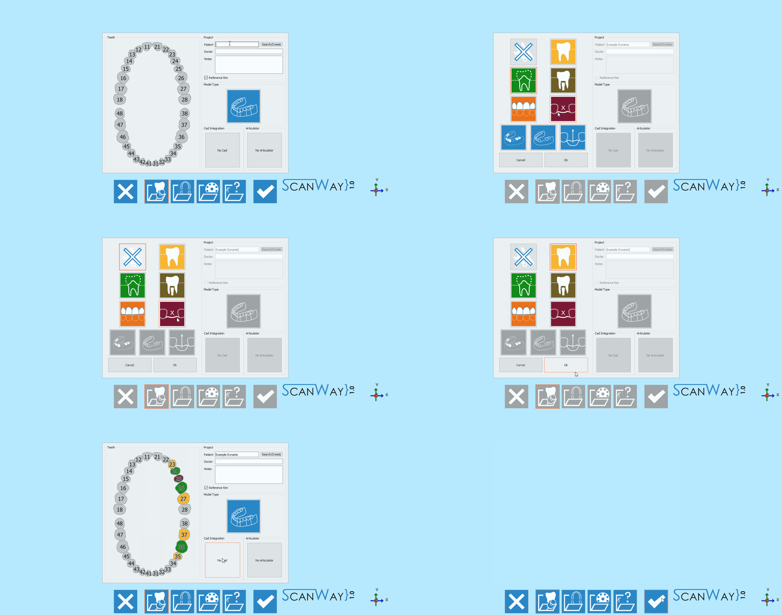
今回のデモ用プロジェクトでは以下の通り定義します。
どちらの方法でプロジェクトを定義しても、同じウィザードに進みます。ここからは、このウィザードに従っていきます。
ステップ1: 咬合器スキャン
両顎を持つように設定されたすべてのプロジェクトの最初のステップは、咬合器スキャンです。
今回のケースでは、咬合器をスキャンします。スキャンされる咬合器と、プロジェクト定義で選択されている咬合器が一致している必要があります。
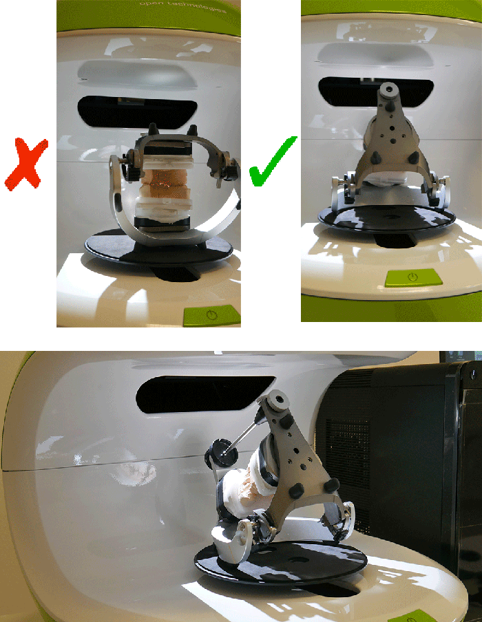
咬合器は後ろに傾けてスキャンすることをおすすめします。これによりできるだけ多くの情報を取得できるようになります。傾けることができない咬合器もあるので、その場合は不要です。
スキャンインターフェースのライブビューでは、咬合器が実際にまっすぐ置かれていると、咬合状態を正確に取得するのが難しくなることが確認できます。
咬合器をスキャナーに設置したら、スキャンボタン![]() でスキャンを開始できます。スキャンが終了すると、結果が表示されます。
でスキャンを開始できます。スキャンが終了すると、結果が表示されます。
利用可能なその他の機能については、スキャンインターフェースのページを参照して下さい。
ステップ2: 下顎
両顎を持つように設定されたすべてのプロジェクトの2つ目のステップは、下顎スキャンです。咬合器を除くすべてのステップには、2つのサブステップ(実際のスキャンと、取得された画像の編集)があります。
スキャンステップ
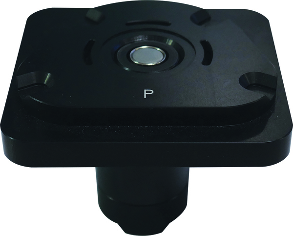
下顎モデルを、選択した咬合器とマッチするスプリットキャストベースに置きます。今回のデモでは、Protarevo- Kavoスプリットキャストベースを使用します。
スキャンボタン![]() をクリックします。スキャンが終了すると、結果が表示されます。
をクリックします。スキャンが終了すると、結果が表示されます。
編集ステップ
このステップでは、取得した画像を編集できます。このステップで利用できるオプションについて詳しくは、編集ツールのページを参照して下さい。
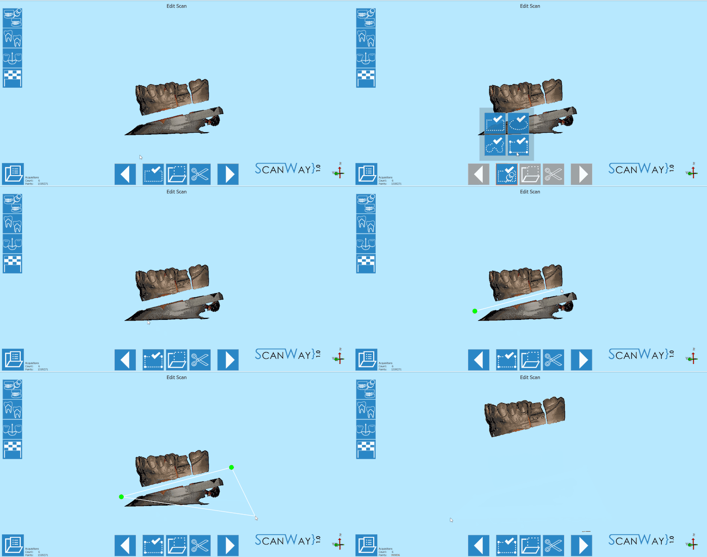
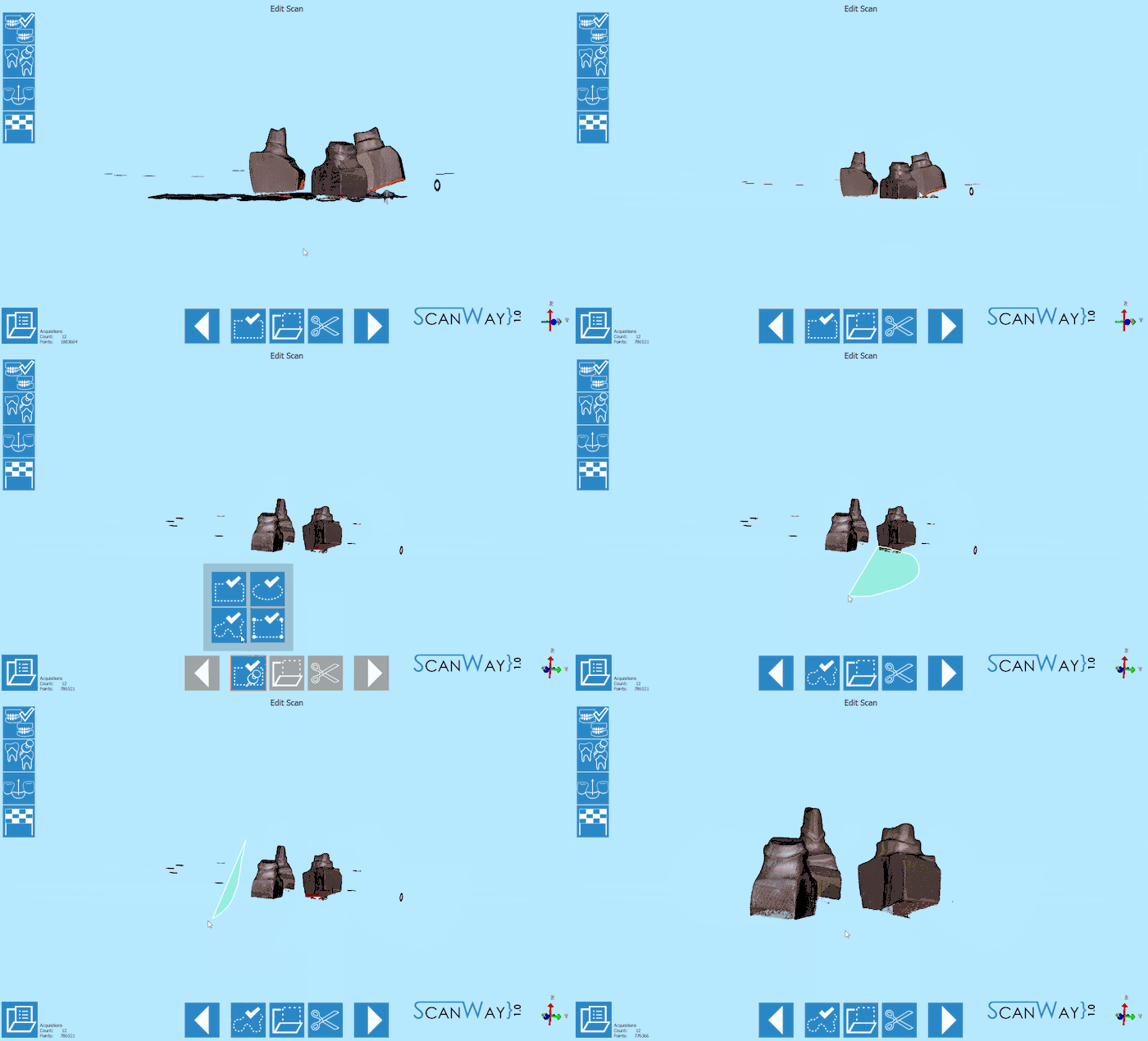
この段階の画像は編集したりトリミングしたりできます。ただし、この段階では画像に対してあまり大きな編集を行ったり、多くの情報を切り取ったりしないことが重要です。このような編集により、オブジェクトと参照モデルの自動アライメントの計算が困難になる場合があります。
今回のケースでは、モデルが咬合器に固定されているので、完全に水平な面でスキャンされません。このため、手早くトリミングするため、多角形選択ツールを使用します。
適切な結果が取得できたら、![]() をクリックして次の手順に進みます。
をクリックして次の手順に進みます。
ステップ3: 上顎モデル
上顎モデルのスキャンでは、下顎モデルと同じように、2つのステップを実行する必要があります。
スキャンステップ
上顎モデルを、選択した咬合器とマッチするスプリットキャストベースに置き、スキャンボタン![]() をクリックします。スキャンが終了すると、結果が表示されます。
をクリックします。スキャンが終了すると、結果が表示されます。
編集ステップ
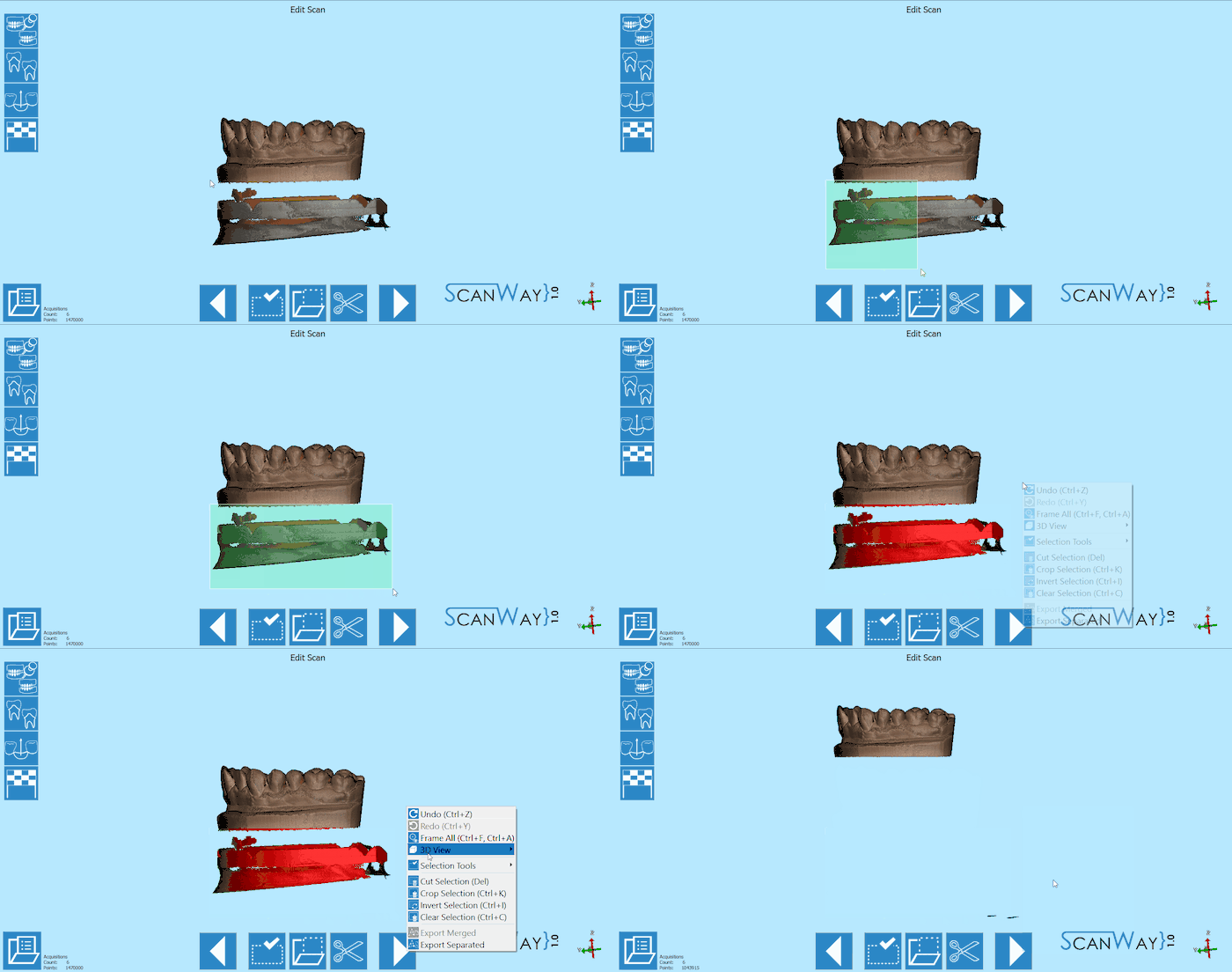
上顎モデルもまだ編集可能です。ここでは、別の選択ツールを使って画像を編集します。
ステップ4: ダイのスキャン
ダイのスキャンに必要なステップは、ユーザーが選択したスキャン方法(マルチダイプレートを使用するか、カスタム設定を使用するか)によって異なります。
マルチダイ使用時
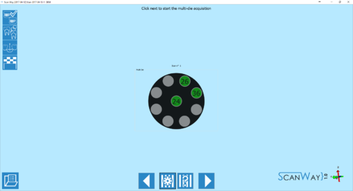
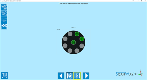
デフォルトではこの方法が適用されています。この場合、マルチダイプレート上でソフトウェアが指示する位置にユーザーがダイを置いていきます。これにより各ダイが直ちに識別されます。
- 定義
- 前述の通り、マルチダイプレートのダイの位置はソフトウェアによって事前に認識されています。
マルチダイの順番は、ユニバーサル方式に基づいており、最初の1/4顎の最初の要素から時計回りの方向に番号が振り当てられています。中央に置く必要のあるダイは、常に最初の1/4顎の最後の要素に最も近いものとなります。
ダイが9個を超える場合、追加の定義ステップが表示されます。
- スキャン
- マルチダイスキャンインターフェースはすでに紹介したほかのステップに似ていますが、相違点として、マルチダイのレファレンスがウインドウ右側のライブビューの下にリマインダーとして表示されます。
続行するには、スキャンボタン をクリックします。スキャンが終了すると、結果が表示されます。
Click ![]() to access the edit step for the multidie scan.
to access the edit step for the multidie scan.
- Edit
- The edit step offers the same tools we saw in previous steps. In this case, more than one tool may need to be applied.
When you are satisfied with the result click ![]() to access the next wizard step.
to access the next wizard step.
- Alignment
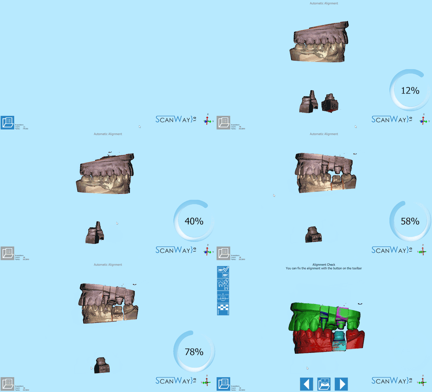
- After being edited the dies get aligned to their references. The result is then showed on the monitor. To learn how to change, fix or redo the alignment check the Alignment Interface page.
Custom Set-Up (Without Multidie)
The user can also decide to scan the dies in a custom order, for instance if he needs to scan the dies on the model base. To access the custom set- up definition, click on the ![]() icon when the software presents the multidie definition.
icon when the software presents the multidie definition.
- Definition
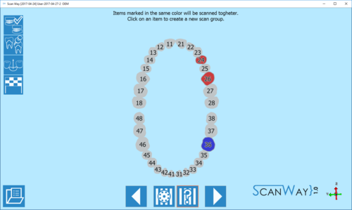
- The software proposes by default a unique scan group which means that, if not defined otherwise, the software will ask the user to scan all the stumps together. In this case I have created a second scan group, to divide the scan of the upper dies from the scan of the lower dies. Click
 to access the scan steps.
- Scan
- Depending on the number of scan groups created, the software will propose one or more scanning steps.
First, the software requires the user to insert the dies in the scanner and acquire the items of the first group.
As a reminder, it marks the items to be scanned together on the right of the window, under the live view.
Then it will ask the user to insert the item selected for the second group. Since the second scan is an individual die, there will be no reminder on the right. Click ![]() to continue.
to continue.
- Dies Identification
- When a scan group has more than one item, the user will be asked to separate each die from the others to correctly identify it.
This step occurs in between the scanning steps. To learn more on the identification of the dies visit the Scan Interface page. Click to continue.
- Edit
- If the scan group is formed by one individual die, there will be no identification step. Instead, the edit step will be presented. Click
 to continue.
- Alignment
- After being edited or identified the dies get aligned to their references. The result is then showed on the monitor. To learn how to change, fix or redo the alignment check the Alignment Interface page.
Step 5: Healthy and Pontics
At this stage the project has been scanned, unless the user wants to rescan the healthy and pontics. This step is infact a result of the scan of the scanned reference model, trimmed to exclude the parts that have been scanned individually (in this case, the dies). It sometimes happens that in the first model scan, the contact points on the healthy are not correctly recognised, which would require the user to rescan the model. Otherwise, just click ![]() to continue.
to continue.
The software will then propose a further edit step to allow the user to modify the healthy image. Proceed in the edit step as previously explained.
Step 6: Mesh Generation and Export
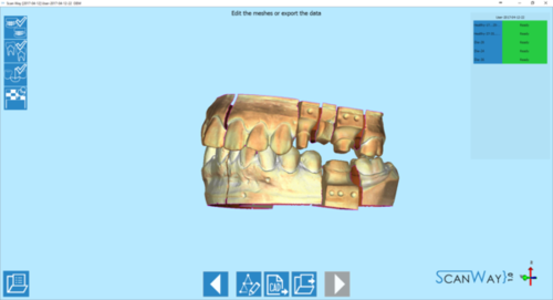
At this point the software immediately starts mesh generation. The meshes can be edited and exported individually or as a unique image. To learn more about mesh editing visit our Mesh Tools page.
Click on the Export Button ![]() to export to CAD.
to export to CAD.
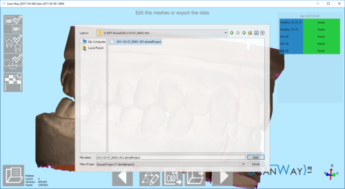
If the project has been started from Exocad, the CAD will automatically open and the design can be started immediately. Otherwise the software will ask the user how to export the file.
Since Exocad has been selected in ScanWay's project definition, the software will ask the user to find the folder containing the DentalProject file for this job.
